
Description
Quick Overview
* Wide area capture including outer peripheral
* Dark area analysis function
* Automatic analysis and a variety of manual analysis modes
* Continuos automatic capturing reduces capturing errors
* Increased speed ensures patient comfort
* Large volume database and back-up capabilities with Sd card
* Overside adjustable touch screen
* Automatic acquisition and automatic shot
Details
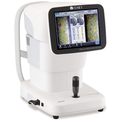
| Wide variety of capturing and analysis functions * Wide Capturing area including peripheral Wide capturing area of 0.25 x 0.54mm can be viewed utilizing original technologies. The endothelium can be viewed in a wide area of the cornea. Having the patient fixate their eye on the fixation light enables the unit to capture images at 15 points in total. The wide range of capturing positions has increased the change of capturing images on patients with partial cornea opacity. A mark indicating the image capture location can be added to the icon that indicates the selected position of the fixation light. Central cornea thickness can be measured simultaneously. The estimated measurement in the ultrasound mode can also be displayed. Smooth and speedy * continuous automatic capturing reduces capture errors Capture errors have been reduced by continuously capturing 16 images with one-time capturing operation. The best quality image is automatically selected and displayed. Selecting the desired image is also possible. |
Reviews
There are no reviews yet.