Description
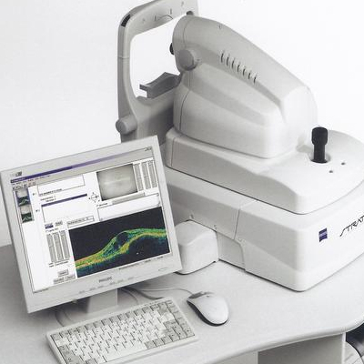
The ZEISS Stratus OCT Model 3000 (Stratus OCT) enables examination of the posterior pole of the eye at an extremely fine spatial scale, without surgical biopsy or even any contact with the eye.
The name Stratus OCT (derived from “stratum,” Latin for “layer”) refers to its unique ability of direct cross-sectional imaging of the layers of the retina. The Stratus OCT minimizes patient discomfort as it permits detailed examination of the retina and optic nerve head at the office or clinic. The Stratus OCT facilitates diagnosis and management of retinal diseases and glaucoma.
Intended Use
The Stratus OCT is intended for use as a diagnostic device to aid in the management of ocular diseases.
Indications for Use
The Stratus OCT is a non-contact, high resolution tomographic and biomicroscopic imaging device. It is indicated for in vivo viewing and axial cross-sectional imaging and measurement of posterior ocular structures, including retina, retinal nerve fiber layer, macula, and optic disc. It is intended for use as a diagnostic device to aid in the detection and management of ocular diseases including, but not limited to, macular holes, cystoid macular edema, diabetic retinopathy, age-related macular degeneration and glaucoma.
Stratus OCT System Described
The Stratus OCT is a computer-assisted precision optical instrument that generates cross sectional images (tomograms) of the retina with ≤ 10 micrometers axial resolution. It works by using an optical measurement technique known as low-coherence interferometry.
How The Stratus OCT Works
The principle of operation of interferometry is analogous to ultrasound, except that it uses light rather than sound. The difference permits measurement of structures and distances on the ≤ 10 micrometer scale, versus the 100-micrometer scale for ultrasound. Another important difference is that optical interferometry does not require contact with the tissue examined, unlike ultrasound.
The Stratus OCT contains an interferometer that resolves retinal structures by measuring the echo delay time of light that is reflected and backscattered from different microstructural features in the retina. The Stratus OCT projects a broad bandwidth near-infrared light beam (820 nm) onto the retina from a super luminescent diode. It then compares the echo time delays of light reflected from the retina with the echo time delays of the same light beam reflected from a reference mirror at known distances. When the Stratus OCT interferometer combines the reflected light pulses from the retina and reference mirror, a phenomenon known as interference occurs. A photodetector detects and measures interference. Although the light reflected from the retina consists of multiple echoes, the distance traveled by various echoes is determined by varying the distance to the reference mirror. This produces a range of time delays of the reference light for comparison.


Reviews
There are no reviews yet.